

Why Do China's Top Class-A Specialists Reject "Photos of Your Films"? Understanding DICOM's Underlying Value
Many patients and family members, when first seeking care abroad, hold their phone up to a window, a lamp or a computer monitor to photograph their films, then send the compressed images over instant-messaging apps. For senior experts in clinical oncology, spinal neurosurgery or complex joint revision, such photos are often "not enough to make a responsible clinical judgment" — and may even hide tiny metastases.
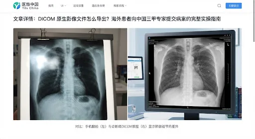
- close The 16-bit grayscale dynamic range is completely lost, crushed down to 8-bit RGB
- close An entire thin-slice CT of 1,000+ layers at 1 mm is reduced to 24 small film-frame photos
- close HU (Hounsfield unit) values and 3D arterial/venous vascular reconstruction become unreadable
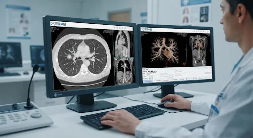
- done Radiologists can instantly switch between lung, bone and mediastinal soft-tissue windows
- done Retains complete spatial data in the axial, sagittal and coronal planes, including multi-phase series
- done Imports directly into MDT review screens and smart radiation-planning systems at China's top cancer hospitals
For an early small lung nodule or a tiny cerebral aneurysm, a 0.625 mm thin-slice scan is what makes minimally invasive biopsy or stereotactic ablation targeting possible. Films usually print only a few dozen selected frames, so micro-lesions are easily missed.
Radiologists need to drag the grayscale: to a bone window to check cortical destruction and micro-fractures, to a mediastinal window to measure the short-axis diameter of lymph nodes. This core capability exists only in the 12–16 bit depth of DICOM data.
For PET-CT, the standardized uptake value (SUV max) of the tracer must be extracted precisely to judge tumor malignancy and response to chemotherapy or targeted therapy — a metric that simply cannot be computed from photos of films.
How to Legally Request and Obtain Your Raw DICOM Data from a Local Medical Facility
Many overseas patients mistakenly believe that the paper report or printed films their doctor gives them are all they get. In fact, whether you are in the US/Europe, Japan/Korea, Southeast Asia or the Middle East, you have a legal right to access and copy your original medical imaging. Most public and private hospitals offer CD/DVD burning or encrypted USB drive transfer services.
folder_zip After You Get the Disc: How to Identify the Master Folder That Holds the Real DICOM Files?
Many hospital discs auto-run a simple "disc browser" viewer when inserted (such as Viewer.exe, Start.exe or Run.app). Please do not send us just this .exe player — executable files usually will not run on another operating system, and they do not contain the images themselves. Instead, open the disc drive and look for the following standard structure:
Lossless Extraction, Packing and Compression on Your Computer (Windows & Mac)
Many modern ultrabooks no longer have a built-in optical drive. You can buy a USB external optical drive for about USD 15–20 on Amazon or at a local electronics store, or borrow one from a nearby print shop. Please follow these four steps carefully:
Step 1: Create a properly named folder on your desktop
Create a new folder on your desktop using English characters only. To prevent files from getting mixed up as they move between multi-specialty consultations, do not use Chinese characters or special characters. Follow this naming scheme:
WANG_XIAOMING_CHEST_CT_20260715 or JOHN_DOE_BRAIN_MRI_20260620 Step 2: Select the entire contents of the disc and copy everything into the new folder
Double-click the disc drive to open its root directory, then press Ctrl + A (Windows) or Cmd + A (Mac) to select all files and folders. Copy them with Ctrl + C and paste with Ctrl + V into the folder you created in Step 1.
Step 3: Pack and compress everything into a standard .ZIP archive
Back on your desktop, right-click the new folder you just filled:
Step 4: A quick 1-minute local self-check — make sure the slices scroll and load properly
To make sure your exported imaging is not corrupted and no key slices are missing, spend 1 minute on a self-check before submitting. We recommend these two globally used, free, medical-grade DICOM viewers:
Simply drag the extracted folder into the RadiAnt window. If series thumbnails appear on the left and the mouse wheel scrolls smoothly through the hundreds of axial slices, your data is intact.
On Mac, Horos is the first choice. Click Import to load the folder, then check that the patient name, study date and the number of series are all displayed correctly.
Submission Priority and Time-Validity Reference Table for Multi-Modality Imaging
When submitting records to different departments of China's top Class-A tertiary hospitals, each specialty weighs imaging modalities very differently. At the same time, medical imaging has a strict "biological time window" of validity. Below are the standardized submission guidelines developed by the MedTravel China expert committee:
| Specialty | Core Required DICOM Modalities | Time-Validity Window | Special Baseline-Comparison Requirements |
|---|---|---|---|
| cardiology Comprehensive Cancer Treatment (CAR-T / Proton) | Multi-phase contrast-enhanced CT of the chest / full abdomen, high-resolution contrast-enhanced MRI of the brain, whole-body 18F-FDG PET-CT | Within 30 days | Original baseline imaging from before chemotherapy / immunotherapy and before surgery is mandatory, to assess response by RECIST 1.1 |
| accessibility_new Orthopedics & Complex Joint Revision | Weight-bearing full-length standing X-ray of both lower limbs, thin-slice 3D CT reconstruction of the affected joint, joint MRI | 60 – 90 days | Patients needing revision of a loose artificial joint should provide the immediate post-operative comparison images taken within the first week after the original replacement |
| water_drop Kidney Disease & Vascular-Access Center | Doppler ultrasound of both kidneys, CT angiography of the renal arteries (CTA), MRI of the retroperitoneal region | 30 – 45 days | Also provide lab reports from the last 7 days: serum creatinine, eGFR and 24-hour urine protein quantitation |
| psychology Neurosurgery / Brain Tumors | 3.0T multimodal brain MRI (T1/T2/FLAIR/DWI/contrast-enhanced), MR spectroscopy (MRS), CTA | 21 – 30 days | Malignant gliomas and brain metastases progress quickly; imaging older than 30 days usually has to be redone as an urgent plain scan immediately after arrival in China |
tips_and_updates Golden Expert Tip: Should You Send the Old Scans at All?
The answer is: absolutely yes! Previous baseline imaging carries as much value for Chinese experts as the newest scans. To tell whether a lung nodule is benign or malignant, for example, comparing thin-slice CT from two years ago and calculating the nodule's volume doubling time (VDT) improves accuracy by more than 40% over a single study. Create a separate ZIP archive for each exam date and upload them all together, in chronological order.
MedTravel China End-to-End Encrypted Cloud Transfer & Data Compliance
Cross-border transfer of patient health information (PHI) is tightly protected by international law. The proprietary "Cloud Imaging Gateway" built by MedTravel China gives patients worldwide a secure, ultra-fast, fully compliant direct link — eliminating the risk of bounced email attachments and privacy leaks through third-party cloud storage.
AES-256 Military-Grade Encryption
Every transfer runs over TLS 1.3 links with AES-256 encryption at rest, certified for cross-border health data under US HIPAA, EU GDPR and China's Personal Information Protection Law (PIPL).
Free Bad-Sector Repair by Certified Technicians
Discs shipped from overseas are often scratched in transit. MedTravel China's dedicated medical imaging engineers run automated slice-integrity checks on every uploaded dataset, rebuild damaged blocks and apply DICOM de-identification.
Direct Delivery to Class-A Hospital PACS
Data is pushed over a dedicated line straight to the diagnostic workstations of experts at our partner Class-A tertiary hospitals in China (such as The First Affiliated Hospital of Sun Yat-sen University and Zhongshan Hospital, Fudan University) — readable immediately with no format conversion.
Upload your ZIP file now through the dedicated encrypted channel — our medical engineers will verify image integrity and quality within 2 hours.
help FAQ — Common Questions from Patients
Q1: My disc holds more than 4 GB of files, and after compressing, the ZIP is still too large to email. What should I do?
A: Never try sending it as an email attachment through a regular mailbox (attachments are easily truncated and lost). Use MedTravel China's dedicated cloud relay channel for extra-large files (supports single files up to 50 GB with resumable upload), or get a private secure endpoint from your 1-on-1 coordinator.
Q2: I had a PET-CT abroad, but the hospital only gave me a paper report and no disc. How do I get one afterwards?
A: Call or email the Radiology Department or the Medical Records Department where the scan was done, provide your Patient ID, and pay a disc fee of about USD 10–25. Hospitals will usually mail the disc by SF Express / DHL or hold it for pickup at the front desk.
Q3: All the files on my disc are pure numeric strings with no extension. Is the disc damaged?
A: No, it is not damaged. PACS systems from GE, Siemens, Philips and other international scanner vendors routinely store DICOM objects under UID strings (such as 1.2.840.113619.2.55...) — these are perfectly valid standard native slices. Never manually add a .jpg extension to them!
Compiled to ISO 13485 medical-device and health-information standards · Copyright MedTravel China